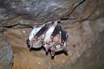
Emerging infectious diseases can become a serious threat to wildlife, a well-known example is white-nose disease, in which the cold-loving fungus Pseudogymnoascus destructans attacks bats during hibernation, penetrating the skin and causing damage. In North America, the disease has already led to the death of millions of bats. P. destructans most likely originates from Europe, where it also causes lesions, but so far it does not seem to have caused any deaths.
However, it is still unclear how the immune system of European bats will deal with this fungal disease in the long term. Even if there are currently no lethal courses of the disease here, the pathogenic fungus could mutate and possibly produce more harmful strains or environmental changes can weaken the bats to such an extent that they show stronger symptoms. It is also still unclear what consequences the disease has, which may not have yet been recognized. Thus, deaths could occur after wintering or there could be negative effects on reproduction.
It is therefore appropriate to monitor infections with P. destructans in Europe as a precautionary measure in the long term, which has also been demanded by EUROBATS for some time (Barova & Streit, 2018). For this purpose, it is important to have instruments that describe the course of the disease exactly. Since hibernation is a physiological condition in which bats are sensitive to disturbances, in-situ assessments of the clinical condition should be performed minimally invasively to avoid adverse effects on bats. However, the two methods for assessing the disease of white-nose disease that have been commonly used worldwide so far required the touch of bats and are therefore invasive: 1) Fluoroscopy of the wing membranes with UV light for the detection of lesions and 2) Quantification of fungal material from wing membrane swabs using qPCR.
After a visual scheme was developed in 2012 to describe the degrees of infestation of the causative agent of white-nose disease on hibernating bats, this methodology was further developed and scientifically verified and referred to as “visual Pd-score”. This non-invasive method is suitable for large-scale monitoring of white-nose disease, as it can be used without disturbing the animals. It is based on a visual classification (scale 0-4), which describes the progress of the infestation (infestation stages) or the clinical condition of the animals. The visual Pd-Score is particularly suitable for routine winter quarters counting or for large-scale P. destructans monitoring. We have prepared a scheme for this purpose, which can be used for the standardized classification (see below).
By the way: Most cases of the infection occur at the end of winter in February and March, when many hibernacula counts have already been completed. It is therefore proposed to carry out appropriate controls at the end of February/beginning of March (mainly hibernation sites of greater mouse-eared bats) and to document the clinical condition of the animals. A detailed article on this topic will appear in the next issue of Nyctalus.
The diagram for determining the clinical condition can already be downloaded here:
Visual assessment of the clinical condition of hibernating bats with regard to white-nosed disease(PDF)

Original studies:
Fritze, M. (2022): Aktueller Wissensstand und langfristige Überwachung der Weißnasenkrankheit bei überwinternden Fledermäusen . Nyctalus 20 (1), accepted.
Fritze, M., S. J. Puechmaille, J. Fickel, G. Á. Czirják, C.C. Voigt (2021): A rapid, in-situ minimally-invasive technique to assess infections with Pseudogymnoascus destructans in bats. Acta Chiropterologica 23, 259–270.
Fritze, M., T. L. H. Pham, B. Ohlendorf (2012): Untersuchung der ökologischen Wachstumsbedingungen des sich auf Fledermäusen ansiedelnden Pilzes Geomyces destructans. Nyctalus 17, 146–160.
